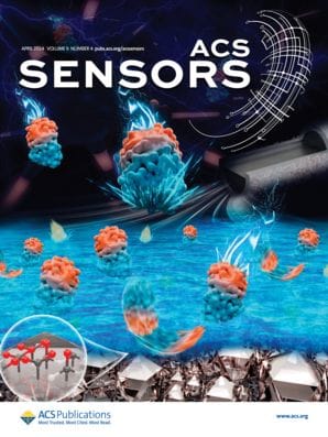
Professor Li-Qun Gu is a Professor of Bioengineering at the Dalton Cardiovascular Research Center at the University of Missouri. He and ACS Sensors Associate Editor Professor Yi-Tao Long of East China University of Science have known each other for more than 15 years. Professor Gu is a pioneer in biological nanopore DNA sequencing while Professor […]

Single-cell exploration at the single-molecule level is essential to understanding heterogeneous cellular functions, such as electron transfer and enzyme activity. Many of these fundamental cellular functions are correlated with the mitochondrial respiration chain. The level of the coenzyme nicotinamide adenine dinucleotide (NADH) is an essential functional indicator of mitochondria.
The assessment of the fundamental cellular processes, such as disease progression and the effectiveness of drugs with minimal destructive, are of great importance. However, the traditional electrochemical method for NADH detection with a micro-scale electrode can damage a living cell, and the fluorescence methods that decrease cell damage will limit precision in NADH detection. Therefore, researchers still need to develop high-spatial- and temporal-resolution approaches for electron-transfer imaging inside a single cell.
Recently, Professor Long and his colleagues developed a glass nanopore electrode to address this challenge, as reported in “Asymmetric Nanopore Electrode-Based Amplification for Electron Transfer Imaging in Live Cells” in the Journal of the American Chemical Society (JACS), which built on his work in Analytical Chemistry, “Wireless Bipolar Nanopore Electrode for Single Small Molecule Detection.” Instead of directly detecting an electrochemical signal, this electrode amplifies the faradaic current produced by electron transfer in a single living cell into a nanopore ionic current signal.
How Researchers Built a Nanopore Electrode
Professor Long and his colleagues built a nanopore electrode using a glass pipette. They reduced the diameter of the pipet tip to tens of nanometers, forming a glass nanopore, then coated the inner surface of the nanopore with a gold layer. By coating the interior of a glass nanopore with a metal layer, a novel wireless electrode was fabricated, achieving the capture of a redox reaction by the ionic current signal read out. Using a nanoelectrode, the NADH level and the drug’s effect were evaluated within a single MCF-7 cell. This method could extend the potential applications of nanopore technology from target single-molecule detection to complex redox process monitoring, which is expected to facilitate the following single living cell analysis to understand the critical cellular processes fully.
The electron transfer of NADH at the electrode’s surface produces hydrogen, which rapidly accumulates to form a “hydrogen bubble.” This bubble, when blocking the nanopore’s ion current pathway, can specifically change the nanopore’s ionic current. The ionic current amplitude is many folds larger than the original electronic transfer current, therefore realizing electronic transfer signal amplification. Using this electrode, the authors can monitor, in real-time, the NADH at concentrations as low as 1 pM.
How Researchers Could Use a Nanopore Electrode
In the past, other researchers developed glass nanopores to measure cellular pH and map the surface charge of single cells. Now, Long and his colleagues have expanded the field by building a nanopore electrode that can monitor real cell electrochemical processes and interpret electron transfer information from inside a single cell.
This novel signal-amplification mechanism, combined with the nanopore’s nanoscale dimension, could be potentially useful in doing high spatial- and temporal-resolution NADH detection inside living cells while minimizing the effects on cellular function.
Long and his colleagues’ findings broaden nanopore technology’s potential applications from target-molecule detection to complex redox-process monitoring. In addition to having a high-current response, the “wireless” electrode has a simplified building process because of the use of the bipolar interface inside the glass nanopore.
This nanoelectrode design may have applications in various single-cell analysis, such as critical cellular processes, cellular communications, and drug screening for cell metabolism regulation, due to its easy modification, controllable nanoscale dimensions, high-sensitivity response, and durable measurement. By modifying with various target probes to the wireless nanopore electrode arrays, the different redox species could be monitored simultaneously. In addition, the small diameter of wireless nanopore electrode provides a high spatial resolution for analyzing intracellular species or processes within a specific organelle.
