A recent study explores a composite bioink for 3D bioprinting, combining a photo-cross-linkable derivative of mucin and hyaluronic acid.
3D printing isn’t just for custom parts for your favorite board game. With the right bio-compatible inks, the technology could revolutionize organ transplants and tissue engineering. In an effort to understand diseases and their progression, teams working in bioengineering are generating physiological models that mimic human tissue, improving our understanding of structural, mechanical, and compositional changes in different tissues and diseases. This is helping to build a 3D picture of different body systems and could one day shape a role for bioprinting in regenerative medicine.1
Mucin is a vital component of mucus, blessed with a certain chemical heterogeneity thanks to its protein backbone and sugar chains and rich in thiol and hydroxyl groups.2 Mucin has many functional groups that can be tailored to suit specific material requirements; it is also rich in STP and cysteine-rich sequences that promote cell attachment. As a result, these regions behave a bit like epidermal growth factor and can play a role in modulating cell growth and proliferation.3
Now, a team working at the Institute of Science in Bangalore, India, have earmarked mucin as a potential bioink for 3D printing.4 The hydrogel nature of mucin makes it a great basis for a bioink, and the researchers chose to pair this with hyaluronic acid—a crucial extracellular matrix component that helps enhance ink viscosity and printability. Hyaluronic acid is naturally found in many tissues and is easy to chemically modify, making it an attractive material platform other applications beyond regenerative medicine, including drug delivery and scaffolds for cell culture.5 The inclusion of hyaluronic acid chimes with other research also looking into its potential as a promising biomaterial.6
In the new study, published in ACS Applied Bio Materials, the team used photo-cross-linking to stabilize the printed scaffolds without damaging cells. The resulting printed structures demonstrated porous behavior conducive to nutrient transport and cell migration. Additionally, after four weeks in a phosphate-buffered saline, the scaffolds retained 70% of their mass, suggesting a good degree of stability.
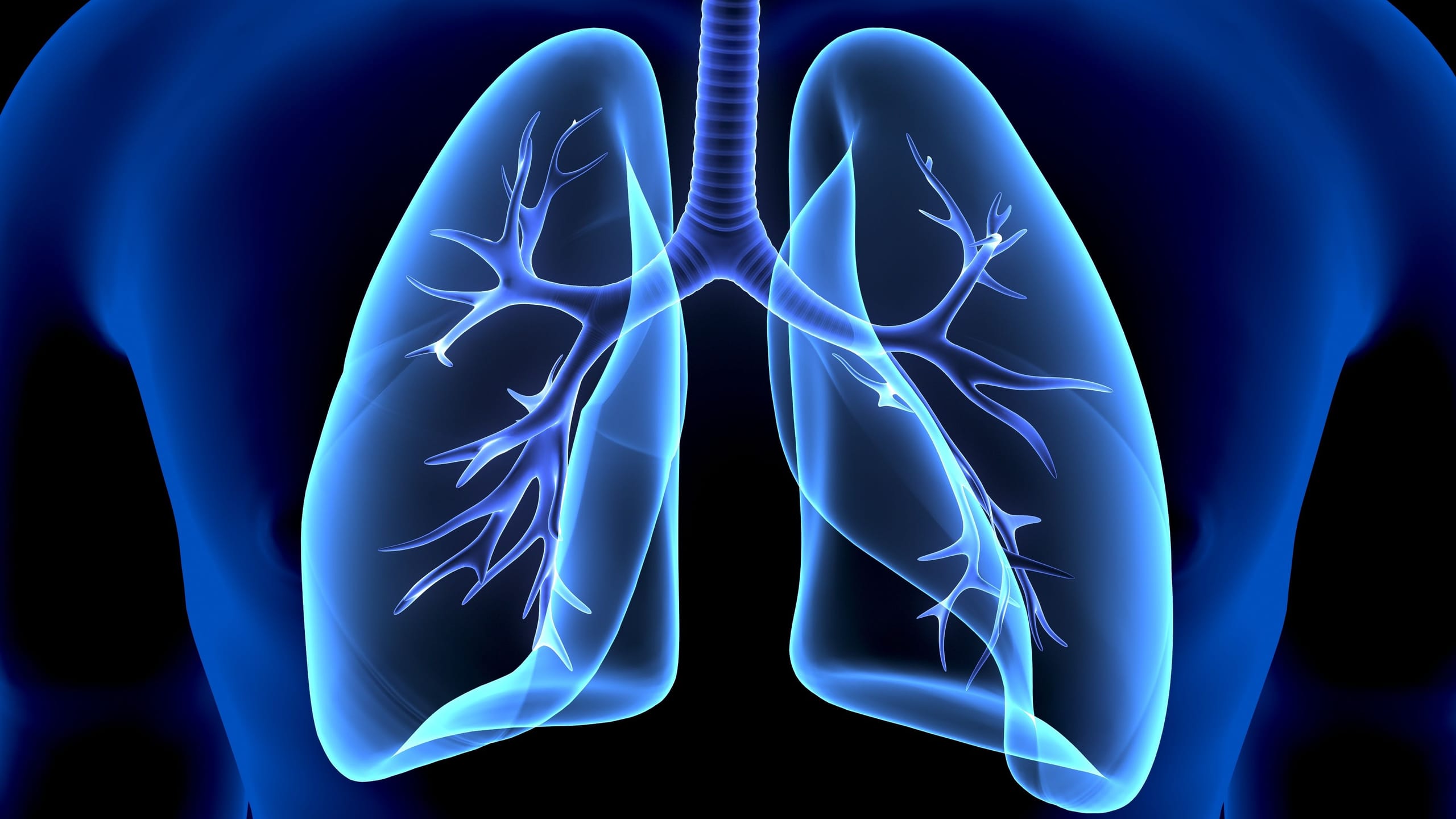
Members of the same team have looked before at using mucin as a repair agent in osteoarthritis or cartilage damage,7,8 but here they were particularly interested in how this new bioink might perform in the lung. Chronic lung disease remains a leading cause of morbidity and mortality, and there is pressing need to deliver new options; this is echoed in a recent Perspective published in ACS Biomaterials Science & Engineering that explores some of the key challenges and opportunities facing the field of lung tissue engineering.9 One of the many challenges in this field surrounds the demand for transplantation—and rising rates of chronic obstructive pulmonary disease, cystic fibrosis, and other respiratory illnesses coupled with donor shortages create a significant gap in this area. This area of research is only around a decade old, founded on reports of engineered rodent lungs that could be implanted orthotopically and provide brief functional gas exchange in vivo.10,11 Since then, a robust infrastructure has been developed, with high-fidelity decellularized extracellular matrix scaffolds being investigated for scale-up to animal and human lungs.12
With this in mind, the new mucin bioink was tested for biocompatibility with lung epithelial cells, with results confirming cell attachment and growth and suggesting suitability for lung tissue engineering. The authors note that the combination of mucin’s versatility and the unique properties of hyaluronic acid makes this composite bioink suitable for a wide range of applications, meriting further investigation in future studies.
References
- Rezaei, F.S. et al. 3D Printed Chitosan/Polycaprolactone Scaffold for Lung Tissue Engineering: Hope to Be Useful for COVID-19 Studies. RSC Adv. 2021, 11 (32), 19508–19520.
- Bansil, R. and Turner, B.S. Mucin Structure, Aggregation, Physiological Functions and Biomedical Applications. Curr. Opin. Colloid Interface Sci. 2006, 11, 164–170.
- Petrou, G. and Crouzier, T. Mucins as Multifunctional Building Blocks of Biomaterials. Biomater. Sci. 2018, 6 (9), 2282–2297
- Sasikumar, S.C. et al. 3D Bioprinting with Visible Light Cross-Linkable Mucin-Hyaluronic Acid Composite Bioink for Lung Tissue Engineering. ACS Appl. Bio Mater. 2024, 7, 8, 5411–5422.
- Später, T. et al. In Vitro and in Vivo Analysis of Adhesive, Anti-Inflammatory, and Proangiogenic Properties of Novel 3D Printed Hyaluronic Acid Glycidyl Methacrylate Hydrogel Scaffolds for Tissue Engineering. ACS Biomater. Sci. Eng. 2020, 6, 10, 5744–5757.
- Wolf, K.J. and Kumar, S. Hyaluronic Acid: Incorporating the Bio into the Material. ACS Biomater. Sci. Eng. 2019, 5, 8, 3753–3765.
- Ajisafe, V.A. and Raichur, A.M. Snail Mucus from Achatina fulica as a Biomaterial Exhibits Pro-Survival Effects on Human Chondrocytes. ACS Biomater. Sci. Eng. 2023, 9, 7, 4208–4222.
- Ajisafe, V.A. and Raichur, A.M. Snail Mucus-Enhanced Adhesion of Human Chondrocytes on 3D Porous Agarose Scaffolds. ACS Appl. Mater. Interfaces 2024, 16, 9, 11324–11335.
- Leiby, K, and Niklason, L.E. Lung Tissue Engineering: Toward a More Deliberate Approach. ACS Biomater. Sci. Eng. 2022, 8, 11, 4625–4628.
- Ott, H.C. et al. Regeneration and orthotopic transplantation of a bioartificial lung. Nature Medicine 2010, 16 (8), 927–933.
- Petersen, T.H. et al. Tissue-Engineered Lungs for in Vivo Implantation. Science 2010, 329 (5991), 538–541.
- Balestrini, J.L. et al. Production of decellularized porcine lung scaffolds for use in tissue engineering. Integr Biol. (Camb) 2015, 7 (12), 1598–1610.