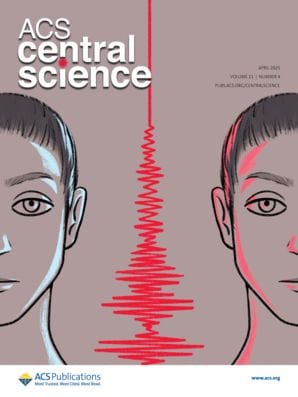
New work explores whether electric-field molecular fingerprinting could overcome current challenges in modern omics and enable simpler workflows for cancer detection.

Our biofluids serve as indicators of various physiological states, and current omics techniques have identified numerous molecular biomarker candidates. However, these are often limited in the number of molecular species they can probe at once, and they often require complicated, target-specific preanalytical workflows.
One alternative approach is molecular fingerprinting, which uses patterns of change across the entire molecular landscape to detect phenotypic changes. Molecular fingerprinting involves leveraging spectroscopic techniques to capture a unique profile of a biological sample (e.g., blood, urine, or tissue). These profiles reflect the presence and abundance of metabolites, proteins, lipids, or nucleic acids, creating a "fingerprint" that can distinguish between healthy and diseased states. If a consistent molecular fingerprint is strongly associated with a specific physiological condition—such as cancer—it can be used to identify or predict that condition, even without knowing the exact biological mechanisms behind it.
In a recent article in ACS Central Science, Mihaela Žigman and colleagues report on a proof-of-concept study using recent developments in laser-based electric-field molecular fingerprinting.1 Previously, the team had worked to develop both a data processing procedure and an in silico model that helped overcome accuracy concerns with molecular fingerprinting while preserving sensitivity.2,3 Additionally, they explored field scaling for trace analyte detection in a 2020 study.4 With new insights gained from their earlier efforts, they wanted to assess the potential of electric-field molecular fingerprinting technology as a platform for in vitro blood plasma profiling for cancer diagnostics. To do this, they collected blood plasma samples from more than 2,500 individuals, spectroscopically profiling bulk venous blood plasma across lung, prostate, breast, and bladder cancer. The researchers used infrared spectroscopy, which examines the vibrational response of molecular bonds to optical excitation by using an ultrashort laser pulse followed by time-resolved sampling of the infrared electric field emitted.5,6

Electric-Field Molecular Fingerprinting to Probe Cancer
With the help of machine learning, the team were able to detect infrared signatures specific to therapy-naı̈ve cancer states, distinguishing them from matched controls. Detection was particularly strong for lung cancer, which could be due to the fact that these tumors often grow faster than other cancers, or perhaps due to the close proximity to the circulatory system. Signals were also shown to increase with disease progression, indicating something of a dose–response effect.
The findings demonstrate that electric-field molecular fingerprinting is a robust framework that could be used for broad disease phenotyping in real-world settings. Of note, each measurement took just 90 seconds and was followed by a cleaning step lasting 2 minutes. “With further technological developments and independent validation in sufficiently powered clinical studies, [laser-based infrared molecular fingerprinting] could establish generalizable applications and translate into clinical practice advancing the way we diagnose and screen for cancer today,” notes Žigman in a recent press release. A First Reactions response to the study also expresses excitement for potential applications while agreeing that the results "justify further efforts to improve the laser system, data acquisition, and analysis methods to...support its use as a minimally invasive screening approach."7
Further Explorations: Related Articles in ACS Journals
Standardized Electric-Field-Resolved Molecular Fingerprinting
Marinus Huber, M. Trubetskov, W. Schweinberger, P. Jacob, M. Zigman, F. Krausz, and I. Pupeza*
DOI: 10.1021/acs.analchem.4c01745
Raman Spectroscopy: A Tool for Molecular Fingerprinting of Brain Cancer
Sivasubramanian Murugappan, Syed A. M. Tofail, and Nanasaheb D. Thorat*
DOI: 10.1021/acsomega.3c01848
Limits and Prospects of Molecular Fingerprinting for Phenotyping Biological Systems Revealed through In Silico Modeling
Tarek Eissa, Kosmas V. Kepesidis, Mihaela Zigman*, and Marinus Huber*
DOI: 10.1021/acs.analchem.2c04711
Molecular Fingerprinting of Mouse Brain Using Ultrabroadband Coherent Anti-Stokes Raman Scattering (CARS) Microspectroscopy Empowered by Multivariate Curve Resolution-Alternating Least Squares (MCR-ALS)
Yusuke Murakami, Masahiro Ando, Ayako Imamura, Ryosuke Oketani, Philippe Leproux, Sakiko Honjoh, and Hideaki Kano*
DOI: 10.1021/cbmi.4c00034
Infrared Spectroscopy for Rapid Triage of Cancer Using Blood Derivatives: A Reality Check
Shaiju S Nazeer*, Ravi Kumar Venkataraman, Ramapurath S. Jayasree, and Jagadeesh Bayry
DOI: 10.1021/acs.analchem.3c02590
References
- Kepesidis, K. et al. Electric-Field Molecular Fingerprinting to Probe Cancer. ACS Cent. Sci. 2025, 11, 4, 560–573.
- Huber, M. et al. Standardized Electric-Field-Resolved Molecular Fingerprinting. Anal. Chem. 2024, 96, 32, 13110–13119.
- Eissa, T. et al. Limits and Prospects of Molecular Fingerprinting for Phenotyping Biological Systems Revealed through In Silico Modeling. Anal. Chem. 2023, 95, 16, 6523–6532.
- Huber, M. et al. Optimum Sample Thickness for Trace Analyte Detection with Field-Resolved Infrared Spectroscopy. Anal. Chem. 2020, 92, 11, 7508–7514.
- Infrared Spectroscopy. ACS Reagent Chemicals: Specifications and Procedures for Reagents and Standard-Grade Reference Materials, online ed., Part 2; American Chemical Society, 2017. DOI: 10.1021/acsreagents.2008
- Baiz, C. et al. Vibrational Spectroscopic Map, Vibrational Spectroscopy, and Intermolecular Interaction. Chem. Rev. 2020, 120, 15, 7152–7218.
- Danotos, M. Detecting the Feeble Electromagnetic Emissions from Cancer Biomarkers. ACS Cent. Sci. 2025, 11, 4, 505–507.
