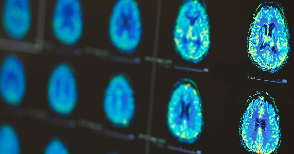
A new study explores a non-invasive imaging technique for measuring the location and concentration of key neurotransmitters in the brain, which could aid in more effective diagnosis and treatment of Alzheimer's disease.

Globally, Alzheimer’s disease is the most common neurodegenerative disorder, affecting millions of people and rising. The number of people living with Alzheimer's disease doubles every five years beyond the age of 65—and in the United States alone, the rates are projected to nearly triple by 2060, potentially affecting around 14 million people.1 Age is a known risk factor, but changes in the brain, such as neurotransmitter levels, can begin many years before the first symptoms appear.
It is common for neurotransmitters to decrease with age, but low levels of adenosine triphosphate (ATP) in particular can be an indication of Alzheimer’s. Most of us will recognize ATP as the major energy currency in cells, but it is also a biomarker for cancer and other diseases.2 The ability to better detect concentrations and cellular locations of ATP in the brain could help increase our understanding about its role in promoting brain health and preventing neurodegeneration, but our ability to selectively detect small molecule metabolites in the brain has been limited. A key factor is the protective nature of the blood–brain barrier (BBB), which prevents sensors from accessing their targets.
To overcome this, a team at the University of Texas at Austin developed a new method for effectively transporting ATP-responsive sensors across the BBB. The researchers developed these fluorescent sensors from pieces of DNA (DNA aptamers) that light up when they bind to a target molecule. The ATP-responsive aptamers were then encapsulated in brain-cell derived microscopic vesicles called exosomes.3 Compared to conventional delivery systems, the loaded exosomes were nearly four times more efficient at passing through the endothelial barrier and releasing the fluorescent sensor into brain cells in a mouse model. The results, published in ACS Central Science, showed unique signal distributions for ATP across different brain regions: healthy mice had significant accumulation in the subiculum and cortex, but in the Alzheimer’s model ATP levels showed depletion in these areas. This suggests the method is capable of determining metabolite location and relative abundance with high spatial resolution in vivo.
DNA aptamers for adenosine and ATP are already showing promise in other imaging and DNA nanotechnology studies, proving to be powerful biosensors.4,5 Another approach being investigated involves new fluorescent dyes that can be used in brain imaging. A 2022 study reports on a series boron difluoride (BF2) formazanate NIR-II dyes, which are very stable, bright, and can be adjusted for different imaging needs. In mouse models, the dyes successfully showed detailed images of the brain and could even distinguish between healthy brain tissue and tumors. These novel avenues of research could one day be used to develop new kinds of non-invasive live brain imaging and potentially expanded to monitor other clinically relevant neurotransmitters.
Related Blog Posts
Beyond Beer—The Preventative Power of Hops
Preventing Alzheimer's Disease With a Daily Shot...of Espresso?
References
- About Alzheimer’s Disease. Centers for Disease Control and Prevention 2024. https://www.cdc.gov.
- Fiorillo, M. et al. High ATP Production Fuels Cancer Drug Resistance and Metastasis: Implications for Mitochondrial ATP Depletion Therapy. Front. Oncol. 2021, 11, 740720.
- Banik, M. et al. Delivering DNA Aptamers Across the Blood–Brain Barrier Reveals Heterogeneous Decreased ATP in Different Brain Regions of Alzheimer’s Disease Mouse Models. ACS Cent. Sci. 2024, 10, 8, 1585–1593.
- Zhang, J. et al. DNA Aptamer-Based Activatable Probes for Photoacoustic Imaging in Living Mice. J. Am. Chem. Soc. 2017, 139, 48, 17225–17228.
- Ding, Y. and Liu, J. Pushing Adenosine and ATP SELEX for DNA Aptamers with Nanomolar Affinity. J. Am. Chem. Soc. 2023, 145, 13, 7540–7547.
- Wang, S. et al. Photostable Small-Molecule NIR-II Fluorescent Scaffolds that Cross the Blood–Brain Barrier for Noninvasive Brain Imaging. J. Am. Chem. Soc. 2022, 144, 51, 23668–23676.

